![]()
![]()
Luciana Cristina Ferretti de Nazareno Wollmann; Carlos A. H Laurindo; Francisco Diniz Affonso da Costa; Andréa Novais Moreno
DOI: 10.5935/1678-9741.20110029
ABSTRACT
The objective of this study was to evaluate the morphology of decelluarized and/or cryopreserved porcine pulmonary valves, to determine a solution capable of completely remove the cells without damaging the extracellular matrix. Porcine pulmonary valves were incubated for 24 hs in sodium deoxicholate 1% or sodium dodecyl sulfate 0.1 and 0.3%, with or without associated cryopreservation. Evaluation was done with optical microscopy (Hematoxilin-Eosin, Acetic Orcein and Gomori) and with morphometric analysis. The effectiviness of the solutions was variable, but the best results were obtained with the sodium dodecyl sulfate solution 0.1%.RESUMO
O objetivo desse estudo foi avaliar a morfologia de valvas pulmonares porcinas criopreservadas e/ou descelularizadas para determinar uma solução que remova as células, sem promover danos à matriz extracelular. Valvas pulmonares porcinas foram incubadas por 24h em soluções de deoxicolato de sódio 1% e de dodecil sulfato de sódio 0,1% e 0,3%, com ou sem criopreservação adicional. A avaliação foi feita com microscopia óptica (hematoxilina eosina, orceína acética ou Gomori) e por morfometria. A efetividade das soluções foi variável, mas os melhores resultados foram obtidos com enxertos frescos descelularizados com dodecil sulfato de sódio 0,1%.INTRODUCTION
Aortic and pulmonary homografts have been used as valve replacement or repair of congenital heart disease for more than 40 years, with satisfactory clinical results [1,2].
Cryopreservation is the method most often used for processing and storage of these grafts, since it preserves the extracellular matrix (ECM) relatively intact with maintenance of cell viability, allowing the storage of the grafts for up to 5-10 years. However, some recent studies suggest that cryopreservation may be associated with structural damage not previously recognized, with significant changes in collagen and elastic fibers and loss of glycosaminoglycans (GAGs) of the ECM [3,4].
It was believed for long that the preservation of cell viability was important, for it would result in grafts with some degree of regenerative capacity and, consequently, with greater durability. However, several studies have shown that after implantation, cryopreserved homografts rapidly become acellular, and subject to progressive tissue degeneration. In addition, there is evidence that high levels of cell viability may even be detrimental, since they arouse more intense immune response by the host, leading to accelerated tissue obliteration [1,3].
More recently, the method of decellularization has been proposed as a promising alternative in the processing of biological tissues [5]. Among the various techniques described are the use of enzymatic treatments [1,6-8], with anionic or nonionic detergents and / or alcohol, either alone or in combination, such as trypsin, Triton X-100, Tween-20, CHAPS (3-[(3-cholamidopropyl) dimethylammonium]-1-propanesulfonate), sodium dodecyl sulfate (SDS), sodium deoxycholate (DOA), glycerol and polyethyleneglycol [9-13].
In theory, the complete removal of the cells results in an acellular matrix and inert the immunological point of view, which can be resettled "in vitro" or in bioreactors "in vivo" by the host, resulting in a graft alive with regenerative capacity and growth [14,15]. The proper preservation of the ECM during decellularization is critical since the future restocking is dependent on the interaction with specific matrix molecules that allow the adhesion, proliferation and cellular differentiation [1,5,16,17].
This study aims to analyze the morphological changes in the ECM from the process of decellularization with different concentrations of SDS or DOA, either alone or in combination with cryopreservation; aiming to determine an optimized tissue processing method for the production of decellularized valve homografts.
METHODS
We used 32 porcine pulmonary valve grafts, which were divided into two groups of 16 grafts each, according to the initial technique of tissue preservation. Group I consisted of fresh grafts, while Group II consisted of cryopreserved grafts. Both groups were divided into four subgroups with four grafts in each, according to the additional treatment of decellularization. Subgroups IA and IIA were decellularized, whereas subgroups I and IIB, I and IIC, and I and IID were decellularized with DOA 1%, SDS 0.1% and 0.3% SDS, respectively. The division of the grafts in the subgroups is shown in Table 1.
Porcine pulmonary valve grafts
We used porcine hearts of Landrace white males, aged between 90 and 120 days. Soon after the slaughter, the hearts were immersed in 0.9% cold saline solution at 4ºC, and then transported to the laboratory. The total ischemic time between the sacrifice and dissection, never exceeded 20 h and the maximum temperature of the transport solution of the graft did not exceed 10ºC. The grafts were prepared by dissecting the muscles of the right ventricle outlet proximally, leaving a rim of muscle just 3 to 5 mm and using a distal transection of the pulmonary artery 1-2 cm above the pulmonary valve commissures. The adventitial layer and the periadventitial fat tissue were carefully dissected to the fullest extent of the arterial graft. Once the dissection and graft preparation was completed, four grafts were immediately placed for histological analysis - Group IA (Control Group - Fresh grafts). All the others were stored for a few hours in cold saline solution and subsequently subjected to the process of cryopreservation and / or decellularization.
Cryopreservation process
For cryopreservation, the grafts were immersed in 100 ml of culture medium RPMI 1640 (Roswell Park Memorial Institute - a mixture of salt fortified with amino acids, vitamins and other components essential for cell growth), 10% of dimethyl sulfoxide (DMSO) and 10% fetal bovine serum (FBS). They were then packed in aluminum pouches sealed and kept at temperature 2-8ºC for 30 minutes. The freezing was done with a tissue cooling speed programmed at -1 to -2ºC/min until the temperature of -80ºC with equipment Planer, KRYO 10 Series, Model 10-16. The grafts were then stored for 15 days in specific containers at a temperature of liquid nitrogen vapor (-195ºC). Thawing was done quickly, with saline solution at temperatures between 42-50 º C, followed by gradual dilution of the cryoprotectant solution with RPMI 1640 and 10% FBS [18].
Decellularization process
For decellularization, the grafts were placed in individual containers containing 100 ml of decellularizing solution (DOA 1%, SDS 0.1% or SDS0.3%), the process being carried out under stirring at room temperature for a period of 24 hours, as PI 0800603-2. After that, the grafts were washed with a solution of sodium chloride 0.9% in agitation for a period of 10 days to ensure complete removal of detergents.
Histological analysis
For histological analysis were obtained 1 cm2 segments of the arterial wall of the channels that were removed from the region just above the sinotubular junction, as well as segments of the central cusp.
Qualitative Analysis: the analysis of tissue morphology was performed with hematoxylin-eosin (HE) and evaluated the presence of cells, the integrity of the ECM and spacing of collagen and elastic fibers.
Quantitative Analysis: we used Gomori trichrome coloring for quantitative analysis of collagen and acetic orcein for elastic fibers. The quantification of collagen and elastic fibers was made with Image-Pro Plus (Media Cybernetics), and captured images of the slides with specific colors for each component matrix (10 images on a 40x objective of each blade in order to capture all the extension of the cusp and random pieces of conduit). Images were photographed with the aid of an Olympus BX50 camera.
Statistical analysis
Morphometric results were analyzed using the Tukey-Kramer multiple comparisons, differences were considered significant with P values <0.05.
RESULTS
Qualitative analysis
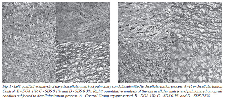
The conduits of the lung control subgroup (IA) had full ECM, with typical pinkish color, well preserved collagen and elastic fibers and cell number and morphology typical of this type of tissue. Conduits decellularized with DOA 1% presented a more compact EMC and more bluish color than the control group. This color can be due to nuclear leakage, since the DNA can fix to the glycoproteins present in the ECM. In addition, there was the presence of several whole cells within this matrix, demonstrating failure of this method to promote complete decellularization. Since the conduits decellularized with SDS 0.1% were completely acellular with no nucleus remains fixed in the matrix, which was visually complete and without compression of the fibers. The conduits decellularized with SDS 0.3% had similar findings, however, one can see discrete spacing of collagen fibers (Figure 1 - left).
The histological appearance of the cryopreserved grafts did not differ much from the fresh grafts, but careful visual observation revealed that the fibers were more distant from each other, giving a looser ECM compared with the "fresh" control. However, it was evident that cryopreservation, when associated with the process of decellularization, significantly worsened the damage in the ECM, especially with the use of the DOA. The conduits cryopreserved and subsequently decellularized with SDS 0.1% or SDS 0.3% proved to be compressed in parts of the conduit and with larger spaces between the fibers, suggesting increased tissue damage (Figure 1 - right).
The analysis of valve cusps showed that all treatments were effective for complete removal of the cells. The cusps treated with SDS 0.1% and DOA 1% were with the ECM looking relatively well preserved, however, treatment with SDS 0.3% resulted in significant damage, with marked compression of the fibers (Figure 2 - left ).
Just as with the conduits, cryopreservation accentuated the damage observed in the ECM of the valve cusps, especially when treated with DOA 1% and SDS 0.3%. The cusps cryopreserved and treated with SDS 0.1%, however, still had relatively well preserved aspect and more similar to the control group (Figure 2 - right).
Quantitative analysis
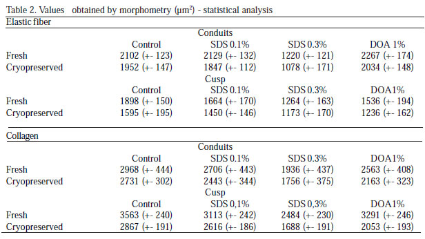
The results found in the morphometric analysis for collagen and elastic fibers are found in Table 2.
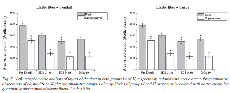
Elastic fibers: Comparing only the grafts in Group I, we find that decellularization with SDS 0.1% and DOA 1% did not significantly reduce the amount of elastic fibers compared to the control group. However, the use of SDS 0.3% resulted in significant differences in the same period (Figure 3).
The grafts in group II had a lower amount of elastic fibers, both in conduct and in the cusps, showing that cryopreservation, per se, interferes with the structural integrity of the fibers. These changes were more significant in the valves decellularized with SDS 0.3%.
Collagen: in the fresh decellularized conduits, the treatments made with SDS 0.1% and DOA 1% do not significantly alter the amount of collagen. The change of the matrix occurs only with the treatment by SDS 0.3% in the fresh ducts. In cryopreserved conduits, there is a significant difference in any of the decellularizing treatments, being the treatment with SDS 0.3% the most important difference. Similar results were found for the cusps (Figure 4).
What is observed in the analysis of fresh grafts is a negligible difference between the control and decellularized groups by treatment with SDS 0.1%, and significant differences in the other two treatments, SDS 0.3% and DOA 1%. In the cryopreserved grafts, there is alteration in all decellularizing treatments, including the cryopreserved cusps without decellularization comparing to the control group.
DISCUSSION
Valve homografts are used for the replacement of native heart valves since 1962 [2]. Since then, various methods of preservation and storage were tested [19-21], however, the technique of cryopreservation; for maintaining the integrity of the ECM, preserving cell viability and allowing storage for long periods of time; has become the methodology more often used by tissue banks.
Currently, it is well established that grafts with cell viability trigger immune reaction by the recipient [1,3,22], however, the correlation between the intensity of this reaction and longevity of the graft is still controversial. Moreover, it is postulated that differences between the techniques of sterilization, processing and storage induce varying degrees of inflammatory reaction, which may adversely affect the longevity of the grafts. The use of acellular matrix, little antigenic and capable of being repopulated after implantation, can, at least in theory, result in graft function and durability [1,5,16].
Ideally, the decellularization technique should be able to completely remove all cells and cell debris and at the same time maintaining the integrity of the extracellular matrix. Our research group at the Center for Cardiovascular Grafts PUCPR tested several decellularizing solutions, and those showing the best results, DOA 1%, SDS 0.1% and SDS 0.3% [8,11,16,17,23,24], were selected for this study. On the other hand, the cryopreservation technique used here was similar to that used by the Banco de Valvas Cardíacas Humanas da Santa Casa de Curitiba [18].
Our results showed that the SDS at both concentrations, was effective in complete elimination of cells of both the cusps and the arterial wall of porcine pulmonary grafts. Since DOA 1% was effective in the decellularization of cusps, however, insufficient to the arterial wall of the conduits. These data demonstrate the importance of adequate concentration and exposure time of the decellularizing solution according to the type of tissue being treated. Thus, a solution that is effective for the decellularization of the pulmonary arterial wall may be insufficient for the decellularization of the aortic wall. On the other hand, the use of more concentrated solutions may be ideal for the wall of the conduit, but at the same time, lead to significant changes in the ECM of the valve cusps.
Histological examination showed that in addition to its better ability to remove cells, SDS promoted less damage to the ECM when compared with the DOA, especially at a concentration of 0.1%. One can also observe that the cryopreservation per se, led to changes in the ECM, and the addition of cryopreservation with the decellularizing solutions enhances the damage. This was most evident in the more delicate tissue of the valve cusps, which were greatly altered when subjected to cryopreservation and later decellularized with DOA 1% or SDS 0.3%.
Controversy exists regarding tissue damage caused during cryopreservation. Schenke-Layland et al. [3] studied fresh and cryopreserved porcine conduits and demonstrated that despite the changes observed by conventional histology are discrete; a more detailed analysis carried out with infrared laser under confocal microscopy can demonstrate ultrastructural deterioration and disintegration of collagen structures. These findings were not confirmed by Gerson et al. [25] who employed the same methodology and showed adequate preservation of the whole structure of the ECM after cryopreservation. Despite the discrepancy of these results not being apparent, differences in the methodology of cryopreservation and of the tissue studied may, at least partially, be concerned.
Literature review suggests that our study is the first to examine changes in collagen and elastic fibers by ECM morphometry. The results demonstrated that decellularization with SDS 0.1% did not alter the amount of elastic and collagen fibers of the cusps and pulmonary conduit when compared to fresh tissue. However, the decellularization with SDS 0.3% or DOA 1% was associated with lower concentrations of these fibers, being these losses significantly marked by the combination of cryopreservation. The results obtained here are unprecedented, since there is no reference to quantitative analysis by morphometric analysis of ECM components in valves subjected to decellularization processes, and they can serve as a reference for future investigations.
The evaluation of decellularized grafts as valve replacements is quite complex, and it involves not only the histological characterization, but its biomechanical characterization, biocompatibility, immunogenicity and potential for cell repopulation. However, it is postulated that a full matrix with a complete absence of cells is essential for obtaining grafts that may be properly repopulated by specific cells, with no tendency to degeneration, showing durability, with regenerative potential and growth. Given the results obtained here, we may suggest that fresh and decellularized grafts with SDS 0.1% solution are the ones that best meet these theoretical premises.
REFERENCES
1. Cebotari S, Mertsching H, Kallenbach K, Kostin S, Repin O, Batrinac A Construction of autologous human heart valves based on an acellular allograft matrix. Circulation. 2002;106(12):I63-8.
2. Ross DN Homograft replacement of the aortic valve. Lancet. 1962;2(7254):487. [MedLine]
3. Schenke-Layland K, Madershahian N, Riemann I, Starcher B, Halbhuber KJ, König K Impact of cryopreservation on extracellular matrix structures of heart valve leaflets. Ann Thorac Surg. 2006;81(3):918-26. [MedLine]
4. Narine K, Ing EC, Cornelissen M, Desomer F, Beele H, Vanlangenhove L Readily available porcine aortic valve matrices for use in tissue valve engineering : Is cryopreservation an option?. Cryobiology. 2006;53(2):169-81. [MedLine]
5. Steinhoff G, Stock U, Karim N, Mertsching H, Timke A, Meliss RR Tissue engineering of pulmonary heart valves on allogenic acellular matrix conduits : in vivo restoration of valve tissue. Circulation. 2000;102(19):III50-5. [MedLine]
6. Tudorache I, Cebotari S, Sturz G, Kirsch L, Hurschler C, Hilfiker A Tissue engineering of heart valves : biomechanical and morphological properties of decellularized heart valves. J Heart Valve Dis. 2007;16(5):567-73.
7. Karim N, Golz K, Bader A The cardiovascular tissue-reactor : a novel device for the engineering of heart valves. Artif Organs. 2006;30(10):809-14. [MedLine]
8. Grauss RW, Hazekamp MG, van Vliet S, Gittenberger-de Groot AC, DeRuiter MC Decellularization of rat aortic valve allografts reduces leaflet destruction and extracellular matrix remodeling. J Thorac Cardiovasc Surg. 2003;126(6):2003-10. [MedLine]
9. Meyer SR, Chiu B, Churchill TA, Zhu L, Lakey JR, Ross DB Comparison of aortic valve allograft decellularization techniques in the rat. J Biomed Mater Res A. 2006;79(2):254-62. [MedLine]
10. Grauss RW, Hazekamp MG, Oppenhuizen F, van Munsteren CJ, Gittenberger-de Groot AC, DeRuiter MC Histological evaluation of decellularised porcine aortic valves : matrix changes due to different decellularisation methods. Eur J Cardiothorac Surg. 2005;27(4):566-71. [MedLine]
11. Booth C, Korossis SA, Wilcox HE, Watterson KG, Kearney JN, Fisher J Tissue engineering of cardiac valve prostheses I : development and histological characterization of an acellular porcine scaffold. J Heart Valve Dis. 2002;11(4):457-62. [MedLine]
12. Mirsadraee S, Wilcox HE, Korossis SA, Kearney JN, Watterson KG, Fisher J Development and characterization of an acellular human pericardial matrix for tissue engineering. Tissue Eng. 2006;12(4):763-73. [MedLine]
13. Kasimir MT, Rieder E, Seebacher G, Silberhumer G, Wolner E, Weigel G Comparison of different decellularization procedures of porcine heart valves. Int J Artif Organs. 2003;26(5):421-7. [MedLine]
14. Schenke-Layland K, Riemann I, Opitz F, König K, Halbhuber KJ, Stock UA Comparative study of cellular and extracellular matrix composition of native and tissue engineered heart valves. Matrix Biol,. 2004;23(2):113-25.
15. Leyh RG, Wilhelmi M, Rebe P, Fischer S, Kofidis T, Haverich A In vivo repopulation of xenogenic and allogenic acellular valve matrix conduits in the pulmonary circulation. Ann Thorac Surg. 2003;75(5):1457-63.
16. Dohmen PM, da Costa FD, da Costa IS, Konertz W Valvas cardíacas obtidas por engenharia de tecidos : A mais nova geração de próteses biológicas. Arq Bras Cardiol. 2002;79(5):555-9. [MedLine]
17. Dohmen PM, Ozaki S, Nitsch R, Yperman J, Flameng W, Konertz W A tissue engineered heart valve implanted in a juvenile sheep model. Med Sci Monit. 2003;9(4):BR97-BR104. [MedLine]
18. Costa MTBA, da Costa FDA, Nazareno LCF, Domachoski J, Peruzzo AM, Colatusso C Análise das atividades dos oito anos iniciais do Banco de Valvas Cardíacas Humanas do Hospital de Caridade da Irmandade da Santa Casa de Misericórdia de Curitiba. Rev Bras Cir Cardiovasc. 2005;20(4):398-407. View article
19. Hopkins RA Historical development of the use of homograft valves1989;:3-13.
20. Hopkins RA Rationale for use of cryopreserved allograft tissues for cardiac reconstructions1989;:15-20.
21. Parker R, Yankah A.C, Yacoub M.H, Hetzer R An international survey of allograft banks : science and practice1997;:5-9.
22. da Costa FD, Dohmen PM, Duarte D, von Glenn C, Lopes SV, Haggi Filho H Immunological and echocardiographic evaluation of decellularized versus cryopreserved allografts during the Ross operation. Eur J Cardiothorac Surg. 2005;27(4):572-8. [MedLine]
23. da Costa FDA, Dohmen P, Lopes SV, Pohl F, Vilani R, Vieira ED Estudo experimental com heteroenxertos valvares descelularizados : a prótese do futuro. Rev Bras Cir Cardiovasc. 2004;19(1):74-82. View article
24. Costa FDA, Dohmen P, Vieira ED, Lopes SV, Colatusso C, Pereira EWL Operação de Ross com homoenxertos valvares descelularizados : resultados de médio prazo. Rev Bras Cir Cardiovasc. 2007;22(4):454-62. [MedLine]
25. Gerson CJ, Goldstein S, Heacox AE Retained structural integrity of collagen and elastin within cryopreserved human heart valve tissue as detected by two-photon laser scanning confocal microscopy. Cryobiology. 2009;59(2):171-9. [MedLine]
Article receive on Saturday, March 12, 2011
 All scientific articles published at bjcvs.org are licensed under a Creative Commons license


All rights reserved 2017 / © 2024 Brazilian Society of Cardiovascular Surgery
DEVELOPMENT BY ![]()
 Read in Portuguese
 Portuguese PDF
 Print
Print
 Send this article by email
 How to cite this article
 Submit a comment
 Mendeley
 Pocket