Article

lock Open Access lock Peer-Reviewed

15

Views

ORIGINAL ARTICLE

Analyses of the immediate effect of CO2 flow on vascular endothelium in goats

Eucário Leite Monteiro ALVESI; Noedir Antonio Groppo StolfII; Ezequiel Cardoso Saraiva de ALMEIDAIII; Luiz Felipe Pinho MoreiraIV; Francisco Solano FEITOSA JÚNIORV; Paulo Sampaio GutierrezVI

DOI: 10.1590/S0102-76382006000300009

ABSTRACT

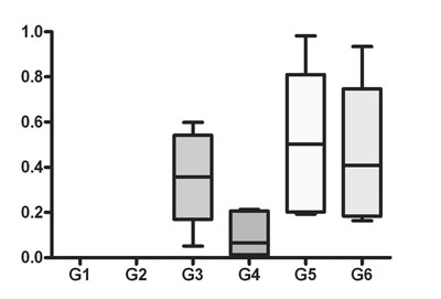
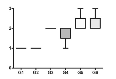
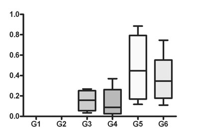
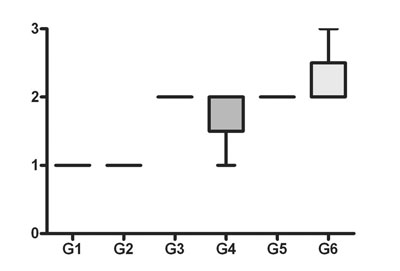
Objective: The purpose of this study was to assess the effect of CO2 on the wall of the Left Internal Thoracic Artery (LITA) and Anterior Interventricular Artery (AIVA) in an experimental model using goats, comparing the immediate effects of the use of CO2 at flow rates of 5 L/min and 10 L/min during 20 minutes, with intermittent flow (every 30 seconds) with and without humidification, simultaneously to the LITA and AIVA. Methods: Thirty-six male goats were submitted to a surgical procedure. Histological analysis was carried out using the immunoperoxidase reaction to mark the endothelium through the detection of VIII Coagulation Factor. Measurement was made by Quantimet following the Ip scale for vascular injury. Results: Within control groups, with and without humidification, both for AIVA and LITA, there was no endothelial injury. The flow rate of 5 L/min provoked moderately significant endothelial injury of the AIVA without humidification, whereas with humidification the endothelial injury was seen but without statistical significance. The flow rate of 5 L/min, with or without humidification, provoked insignificant endothelial injury at LITA. With a flow rate of 10 L/min, there was highly significant endothelial injury, both for the LITA and AIVA and whether humidified or not. Conclusions: In conclusion endothelial injury is flow-dependent with greater injury when using CO2 at a flow rate of 10 L/min and less at 5 L/min. The arteries involved in anastomosis (LITA and AIVA) are both affected, but there is a greater effect on the AIVA.

RESUMO

Objetivo: O objetivo deste estudo é avaliar a influência do jato de CO2 na parede da Artéria Torácica Interna Esquerda (ATIE) e Artéria Interventricular Anterior (AIVA), em um modelo experimental em caprinos, comparando os efeitos imediatos do uso do CO2 com velocidade de fluxo de 5 L/min e 10 L/min, por 20 minutos, com fluxo intermitente (30 em 30 segundos), com e sem umidificação, simultaneamente nas ATIE e AIVA. Método: Trinta e seis caprinos foram submetidos ao procedimento cirúrgico. A análise histológica foi feita por meio da reação de imunoperoxidase para marcação do endotélio, por meio da detecção do Fator VIII da coagulação. A mensuração foi feita pelo Quantimet, seguindo a escala de Ip para lesão vascular. Resultado: Nos grupos controle, com e sem umidificação, não houve lesão endotelial. A velocidade de fluxo de 5 L/min provocou lesão endotelial com nível de significância intermediária na AIVA, sem umidificação; e, com a umidificação, a lesão ocorreu, porém, sem significância. A velocidade de fluxo de 5L/min, com e sem umidificação, provocou lesão endotelial na ATIE, contudo, sem significância. Com a velocidade de fluxo de 10 L/min, houve lesão endotelial com nível de significância importante, tanto para a ATIE quanto para a AIVA, não alterando se umidificado ou não. Conclusão: Pôde-se concluir que a lesão endotelial é fluxo-dependente. As artérias envolvidas na anastomose (ATIE e AIVA) foram afetadas simultaneamente, sendo que a agressão foi maior para AIVA .
INTRODUCTION

An accurate surgical technique is extremely important for a successful surgical result in off-pump coronary artery bypass grafting. Several resources are applied during this kind of intervention to obtain better visibility of the surgical field, including frequent swabbing of arteriotomies using absorbent materials (gauze), the use of stabilizers, intermittent irrigation using saline solution, occlusion of the coronary artery using sutures or clamps (Bulldog), intraluminal coronary shunt and the use of high flows of gas (oxygen, carbon dioxide and compressed air) directed at the anastomoses. However, each technique has its limitations including the risk of endothelial lesion caused by the use of absorbent materials, stabilizers, temporary occlusion of the coronary artery or intraluminal coronary shunt and hemodilution or difficulty to see the surgical field due to intermittent irrigation with saline solution [1].

The use of high flows of gas directed at the anastomosis improves vision of the surgical field and maintains the artery 'ready', aiding the surgical technique [2,3]. Complications, such as coronary air embolism due to the use of O2 and compressed air, justify the preference for CO2, which is 34 times more soluble than air in water at 30ºC [4].

There are few studies about the deleterious effect of CO2 jet on the vascular endothelium. Burfeind et al. [1] reported that a CO2 jet of 15 L/min directed at the anterior interventricular artery of pigs caused endothelial injury with exposure of blood elements to the sub-endothelium, leading to blood clots and endothelial loss which may cause migration and cellular proliferation to the smooth muscle. Okazaki et al. [5] studied the endothelial lesion in dogs during off-pump coronary artery surgery with coronary clamping and CO2 insufflation at 5 L/min and exposure of the first diagonal branch to 'non-humidified' CO2, CO2 'humidified' with Ringer lactate solution and CO2 'humidified' with heparin and dipyridamol associated to Ringer lactate, with a continuous exposure of 10 and 20 minutes. The authors verified lesions in the "non-humidified" group with less in the "humidified" groups. No study was performed using two grafts related to an anastomosis.

Controversies regarding the deleterious effects of CO2, as a method to improve the surgical field conditions, justify further studies.

METHOD

All animals used in the study were handled according to norms established in the 'Guide for the Care and Use of Laboratory Animals' (Institute of Laboratory Animals Resources, National Academy of Science, Washington, D.C., 1996) and by the Ethics Principles established for animal experimentation by the Brazilian College on Animal Experimentation - COBEA (1991). The study project was submitted to the Ethics Commission of the Hospital Veterinário Universitário Dr. Jeremias Pereira da Silva, from Centro de Ciências Agrárias - Federal University of Piauí, where the experimental surgeries were performed, and also to the Ethics Commission of the Hospital das Clínicas Medical School of the University of São Paulo (HCFMUSP).

The study model is primary, experimental, laboratorial, controlled and randomized with double blind analysis of the glass slides.

Division of the groups

Six groups of six goats were formed according to these categories: G1- Control group without the use of "non-humidified" CO2; G2 - Control Group without the use of "humidified" CO2; G3 - Experimental Group with a 5 L/min blow rate of "non-humidified" CO2; G4 - Experimental Group with a 5 L/min blow rate of "humidified" CO2; G5 - Experimental Group with a 10 L/min blow rate of "non-humidified" CO2 and G6 - Experimental Group with a 10 L/min blow rate of "humidified" CO2.

Surgical technique

Thirty-six non-castrated clinically healthy male Anglo-Nubian goats of approximately 18 months old and weighing from 13 to 15 kg were submitted to pre-anesthetic medication (PAM) using 2% acepromazine at a dose of 0.25 mg/kg. At the end of the latency period of pre-anesthetic medication, each animal had its left brachial vein punctured with a 20 Jelco catheter to infuse medicine and the Invasive Mean Arterial Pressure (IMAP) was measured by catheterization of the left auricular artery with another 20 Jelco catheter connecting it to an extension tube, two three-way taps, washed in heparinized saline solution (1 mL in 500 mL of 0.9% SF), with all the system connected to an aneroid barometer. Anesthetic induction was performed 30 minutes after the PAM, with an association 3 mg/kg of 5% Ketamine, and 0.2 mg/kg of Midazolam. Subsequently, the animal was intubated with a number 7 orotracheal tube that was connected to a volatile anesthetic device (Takaoka - Nissei Takaoka Brazil), with a bivalved circular-type circuit and inhalant anesthesia was initiated using Halotane. Hemodynamic control was carried out using IMAP and control of the heart rate and respiratory frequency. Dopamine was administered at a dose of 5-8 mcg/kg/min (mean 7.5 mcg/kg/min) when necessary.

When the animal was under anesthesia, anti-sepsis of the lateral and anterior ventral thoracic region was performed using topical povidine and sterile surgical drapes were placed following conventional criteria. Thus, incision of the skin, thoracotomy and transverse sternotomy were carried out to expose the mediastinum and pericardium, followed by sternal periosteum hemostasis and longitudinal pericardiotomy. Dissection of the pedicled Left Internal Thoracic Artery (LITA) was achieved. Distal ligature near to the LITA bifurcation was performed after heparinization at a dose of 1 mg/kg, to prepare the vascular bed for anastomosis.

Protocol

All animals were submitted to the same protocol, in which after measuring the LITA over the heart to better adapt the space to be used, two intramyocardial sutures using 4.0 prolene thread in a form of proximal and distal tourniquets, with approximately two centimeters between them, were made on the distal bed of the Anterior Interventricular Artery (AIVA), occluding the vessel and establishing the surgical field for the procedure. After obstruction of the AIVA at the determined points, a lengthwise opening of the vessel of approximately two centimeters was made. The distal bed of the LITA was anastomosed to the AIVA using three single sutures (proximal, medium and distal) of 7.0 prolene thread, which were unilaterally equidistant. Exposure of the vessel lumen followed pre-established criteria, according to the speed of the gas flow at 5 or 10 L/min and the presence or absence of humidification.

For the "non-humidified" group, exposure of the vessel lumen occurred for 20 minutes in normal air, without any substance that could possibly humidify the endothelium. For the "humidified" group, small quantities of blood from the LITA were applied at 30-second intervals to lubricate the lumen of the exposed vessels for 20 minutes.

Exposure of the vascular bed to a CO2 jet occurred using a specific catheter manufactured by the Fundação Adib Jatene - SP. The variation in the flow rate in this experiment was 5 and 10 L/min at 4 to 6 centimeter from the anastomosis and at 30-second intervals.

Sacrifice

Subsequently the exposed segments of AIVA and LITA were sectioned and placed in containers with 10% buffered formaldehyde and the animals were sacrificed with a lethal dose of potassium. The Pathologic Anatomy Service of INCOR-HCFMUSP analyzed the segments.

Material analysis

The material was stained using the hematoxylin-eosin (HE) method to evaluate the general aspect of the material. Verhoeff stain was applied to identify and measure the internal elastic lamina and the immunoperoxidase reaction was used to mark the endothelium by coagulation factor VIII detection. Histological analysis of the material was performed by light microscopy at 20x magnification and transferred to a Quantimet, Leica QWIN Standard image analyzer for manual and automatic measurements, including filters of structures according to form, color and size. The measurements of the vascular bed, endothelium and internal elastic lamina were performed in pixels, the size of which is 0.97µ.

Immunoperoxidase technique

Histological sections, following those used for HE staining, were submitted to the immunoperoxidase reaction to detect the human antigen linked to Von Willebrand Factor VIII.

Deparaffinization was performed at room temperature, with three substitutions of xylene at 15-minute intervals, followed by hydration in alcohol at decreasing concentrations (100º, 95º and 70º alcohol) until rinsing in water. Subsequently, endogenous peroxidase was blocked with a 3% hydrogen peroxide solution in a phosphate buffer saline (pH=7.4) with three incubations of 10 minutes each in a water bath at 37ºC.

Subsequently, glass slides were submitted to antigen retrieval by heat, incubating the material in a Tris/EDTA buffer (Hydroxymethylaminomethane /Ethylenediamine Tetraacetic Acid) at pH=9.0 and Tween 20 detergent in a water bath at 95°C for 25 minutes and subsequent cooling at room temperature for 20 minutes; they were rinsed three times in a phosphate buffered solution (PBS) at pH=7.3. The glass slides were incubated with 1% fetal bovine serum at 37°C for one hour to block unspecific proteins.

The polyclonal primary antibody, A0082, produced in rabbits (Dako Cytromation, Carpinteria, CA, USA) was used to detect the human antigen related to Factor VIII at a dilution of 1:500 at 4°C for 18 hours.

The secondary antibody used in the experiment was the rabbit anti-immunoglobulin produced in pigs, conjugated with biotine (Dako Cytromation, Carpinteria, CA, USA) at a dilution of 1:800 at 37°C for 1 hour. The streptoavidine conjugated with peroxidase (Dako Cytromation Carpinteria, CA, USA) was used as the ligation compound for biotine at a dilution of 1:600 at 37°C for 30 minutes.

The glass slides were placed for 1-5 minutes in a chromogenous solution of DAB (3,3'-diaminobenzidine - Dako Cytromation, Carpinteria, CA, USA), after counter-stained with Harris hematoxylin, which stains the nuclei in blue and then they were mounted using cover slips with Entellan resin (Merck, Germany) to see the antigen-antibody reaction.

Classification criteria of endothelial lesions

As the vascular lesion represents the initial event of pathogenesis in several vascular diseases, including atherosclerosis, Ip et al. [6] proposed a morphological and functional classification of the vascular lesion:

 TYPE 1 - There is a functional alteration of the endothelial cell without significant morphological changes;

 TYPE 2 - There is endothelial denudation and intimal lesion, but the internal elastic laminas and media are intact;

 TYPE 3 - There is endothelial denudation with both intimal and media lesions.

Definition criteria of endothelial lesion, bed and internal elastic lamina

The presence of endothelial cells on the vascular bed with brownish coloration due to the immunoperoxidase reaction was considered normal.

The absence (denudation), erosion or delamination of endothelial cells on the vascular bed was considered endothelial lesion (Figure 1).


Fig. 1 - Light photomicrography at 20x magnification of the Anterior Interventricular Artery vascular bed, with a "non-humidified" 5 L/min CO2 blow rate. Immunoperoxidase reaction to mark the endothelium by detection of the coagulation factor VIII


The internal elastic lamina, analyzed after Verhoeff staining, was tortuous along the entire extension of the vascular bed. The AIVA presents with only one internal elastic lamina, while the LITA has several elastic laminas. The discontinuity of the elastic lamina in its extension was considered a lesion.

Discontinuity or erosion of the vascular bed was considered a lesion. There was always extreme caution to differentiate lesions of the bed, with technical problems occurring during the preparation of the glass slides using immunoperoxidase.

Statistical method

Statistical analysis was achieved using the Kruskal-Wallis test for differences among medians, which is a non-parametric test to compare continuous and unpaired data in different groups, with p-values < 0.0001 considered significant. The Kruskal-Wallis test was complemented by Dunn's test for multiple comparisons and the differences were considered significant with p-values < 0.05 and non-significant (ns) when the p-values > 0.05, an intermediate level of significance (*) with p-values < 0.05 and an important level of significance (**) when the p-values < 0.01.

RESULTS

There were no conditions to statistically measure the lesions of the vascular bed and internal elastic lamina, both for the AIVA and LITA, as the results were not significant. The following analysis is related to vascular endothelium injuries.

1- Anterior interventricular artery

1.1 Analysis of AIVA endothelial lesion as a percentage

The Kruskal-Wallis test for AIVA endothelial injuries as percentages detected a difference in median values among groups H=27.86 with a p-value < 0.0001.

The median, maximum and minimum or separatrix values, 1st and 3rd quartiles are presented in Figure 2.


Fig. 2 - Endothelial injury of the Anterior Interventricular Artery as a percentage among groups


There was no significance for G1 and G2. G4 had the lowest median and variability with almost no cases above the 3rd or under the 1st quartile. G5 had the highest variability among the groups. G3 presented intermediate variability, higher than G4 and lower than G5 and G6.

The comparison of G1 ("non-humidified" control) with G5 ("non-humidified" 10 L/min flow) and G2 ("humidified" control) with G5 ("non-humidified" 10 L/min flow) showed endothelial lesion with important significance as seen by Dunn's test. The intermediate level of significance occurred when G1 ("non-humidified" control) was compared with G3 ("non-humidified" 5 L/min flow) and G6 ("humidified" 10 L/min flow) and when G2 ("humidified" control) was compared with G3 ("non-humidified" 5 L/min flow) and G6 ("humidified" 10 L/min flow). The other comparisons among groups were not significant.

1.2 Ip scales for the anterior interventricular artery

The Kruskal-Wallis test for Ip scale in AIVA detected a difference in median values among groups H=29.49 with a p-value < 0.0001.

G1 and G2 showed a uniform trend, with all cases in scale 1 without variations. G3 presented all cases in scale 2 without variations. G4 had a minimum value of 1 and 75% in scale 2. G5 and G6 showed uniformity with a maximum value of 3 and 75% in scale 2 (Figure 3).


Fig. 3 - Ip index for the Anterior Interventricular Artery among groups


The results of Dunn's test for multiple comparisons showed endothelial lesions with an important significance level when G1 ("non-humidified" control) was compared with G5 ("non-humidified" 10 L/min flow) and G6 ("humidified" 10 L/min flow); and when G2 ("humidified" control) was compared with G5 ("non-humidified" 10 L/min flow) and G6 ("humidified" 10 L/min flow). The level of significance was intermediate when G1 ("non-humidified" control) was compared with G3 ("non-humidified" 5 L/min flow) and when G2 ("humidified" control) with G3 ("non-humidified" 5 L/min flow). The other comparisons among groups were not significant.

2-Left internal thoracic artery

2.1 Analysis of LITA endothelial lesion in percentage

The Kruskal-Wallis test for LITA endothelial lesion in percentages detected a difference in median values among groups H=27.42 with p-value < 0.0001.

There was no significance for G1 and G2. G4 had the lowest median and variability higher than G3. G5 had the highest variability among the groups, followed by G6 (Figure 4).


Fig. 4 - Endothelial lesion of the Left Internal Thoracic Artery as a percentage among groups


In the analysis of Dunn's test for multiple comparisons, endothelial lesion had an important significance level when G1 ("non-humidified" control) was compared to G5 ("non-humidified" 10 L/min flow) and to G6 ("humidified" 10 L/min flow); and when G2 ("humidified" control) was compared to G5 ("non-humidified" 10 L/min flow) and to G6 ("humidified" 10 L/min flow). The other comparisons among the groups were not significant.

2.2 Ip Scale for the left internal thoracic aorta.

The Kruskal-Wallis test for Ip scale for the LITA detected a difference in median values among groups H=30.46 with a p-value < 0.0001.

G1 and G2 showed a uniform trend, with all cases in scale 1 without variations. G5 and G6 also showed uniformity with all cases in scale 2, without variations. G4 had minimum value of 1 with 75% in scale 2. G6 presented maximum value of 3 with 75% in scale 2 (Figure 5).


Fig. 5 - Ip index for the Left Internal Thoracic Artery among groups


The results of Dunn's test for multiple comparisons showed endothelial lesion with an important significance level when G1 ("non-humidified" control) was compared with G6 ("humidified" 10 L/min flow) and G2 ("humidified" control) and when G2 ("humidified" control) was compared with G6 ("humidified" 10 L/min flow); The level of significance was intermediate when G1 ("non-humidified" control) was compared with G3 ("non-humidified" 5 L/min flow) and G5 ("non-humidified" 10 L/min flow); and when G2 ("humidified" control) was compared with G3 ("non-humidified" 5 L/min flow) and G5 ("non-humidified" 10 L/min flow). The other comparisons among groups were not significant.

DISCUSSION

The search for a surgical procedure capable of reducing the damage of coronary insufficiency is old, as although clinical treatment can prolong and improve the patient's quality of life, it does not modify the natural history of coronary occlusion. Coronary artery bypass surgery is the most frequent heart surgery performed in Brazil.

With the use of cardiopulmonary bypass (CPB) there are some complications including systemic inflammatory response syndrome [7,8]. Thus, there was a necessity to perform off-pump coronary artery bypass surgeries. However, it is good to remember that the first experiments did not use CPB [9,10]. Reports on off-pump coronary artery bypass surgeries with good results promoted greater use of this surgical procedure [11,12].

On this aspect, many ways to assist off-pump coronary surgeries appeared with the aim of producing satisfactory results [13,14]. Carter & Roth [15] performed an experiment to graft the LITA to the circumflex artery in dogs using an intraluminal metallic ring connected to LITA.

Bleeding caused during arteriotomy encouraged some surgeons to look for techniques to clean the surgical field, including Rivetti et al. [16] who developed an intraluminal shunt (temporary intraluminal shunt), which was introduced into the lumen of the vessel during the anastomosis of the graft to the coronary. Buffolo et al. [14] employed occlusion of the coronary flow using small silicon tourniquets and a distal perfusion system to the occluded artery.

Poirer et al. [17] reported the use of stabilizers on the myocardium wall to cause a smaller degree of intraluminal stenosis in LITA and AIVA anastomoses, during off-pump coronary artery bypass surgery.

Sokullu et al. [18], through studies performed in dogs using electronic scanning microscopy and light microscopy, observed the presence of endothelial injuries with the use of 4.0 prolene thread and bulldog tourniquets. The latter produced less injury.

Other techniques such as frequently swabbing the arteriotomy with absorbent materials (gauze) and intermittent irrigation with saline solution have also been reported [1].

The idea of using gas jets initiated when Sawyer et al. [19] reported the use of a 15 L/min CO2 jet to perform an endarterectomy as a way to improve the visibility of the surgical field. Subsequently, Teoh et al. [2] reported the safe use of oxygen at a 12 L/min blow rate (mean from 10 to 15 L/min). Maddaus et al., [3] reported no complications after using compressed air at a 12 L/min blow rate (mean 10 to 15 L/min), "humidified" with saline solution (1 to 5 mL/min). On the other hand, these data were contested by Sasaguri et al. [4] who reported the risk of coronary air embolism with the use of oxygen and compressed air and proposed the use of CO2 due to its high solubility in the blood.

The use of CO2 for clearing the surgical field and to assist anastomosis has been routinely used by many surgeons, but there are few reports on the effects of CO2 use in the endothelium. Thus, Burfeind et al. [1] performed median sternotomy in seven pigs to expose their anterior interventricular artery to a 15 L/min CO2 jet without humidification for 20 minutes and analyzed the vessel by immunohistochemistry. The result demonstrated a type 2 lesion on the IP scale, with endothelial desquamation that varied from 50 to 100% of the vascular circumference.

Chavanon et al. [20], using an experimental model in pigs, compared several haemostatic models used to revascularize the myocardium without CPB. The experiments demonstrated that clamping produced significant endothelial dysfunction, greater than the use of a gas jet and also the use of the intraluminal perfusion catheter with silicon and Anastaflo caused significant endothelial dysfunction, greater than extravascular occlusion techniques.

Okazaki et al. [5] reported endothelial lesion during off-pump coronary artery surgery in dogs, with coronary clamping and CO2 insufflation at a 5 L/min blow rate. Exposure of the artery was by left lateral thoracotomy. The target vessel was the first diagonal branch which was continuously exposed for 10 to 20 minutes to non-humidified CO2, humidified CO2 with Ringer lactate and humidified CO2 with heparin and dipyridamol associated with Ringer lactate. An investigation of the coronary endothelium was performed by electronic scanning microscopy, which evidenced injury in the non-humidified group and less in the humidified group. The presence of endothelial lesions occurred with exposure to CO2 flows over 20 minutes and decreased with a reduction of exposure time. There was a reduction in endothelial lesion in the humidified group, with a slight difference favoring the group treated with heparin and dipyridamol.

In this study, an experimental model similar to clinical conditions was created involving the conventional arteries, AIVA and LITA, with quantification in extent and percentage of endothelial lesion. With the comparison of different blow rates, it was possible to gain a better notion of the damages provoked in the endothelium. Our choice for a 10 L/min blow rate was similar to the rate used by Teoh et al. [2], who used a 12 L/min oxygen blow rate (mean 10 to 15 L/min) for the first time, which is slower than the 15 L/min blow rate used by Burfeind et al. [1]. The use of a 5 L/min blow rate is closer to the reality of every-day use.

Another parameter chosen was humidification and non-humidification with blood as the proposed vehicle, which physiologically with anticoagulation does not cause damage to the endothelium and which also is the motivational factor for the use of CO2 jet on the vascular bed.

During AIVA occlusion, we applied a tourniquet using intramyocardium sutures of 4.0 prolene thread in an area of myocardium segmentation irrigated by the vessels, causing cyanotic coloration, suggesting immediate ischemia of the area. This fact differs for humans, possibly because goats have less intra or intercoronary collateral circulation. Hence, this fact opens up the wide possibility of studies on not only the heart, but the kidneys, liver and lungs which is a segmented organ.

During the pilot study, the occlusion of the proximal bed caused a greater hemodynamic instability in the animal, with a large ischemic area which consequently caused ventricular fibrillation. In the area exposed to the CO2 jet, when the septal or diagonal branch ostia were exposed, the gas penetrated the vessels, mostly at a 10 L/min blow rate, leading to ventricular fibrillation. For this, the distal bed of the AIVA between the diagonal branches was used in the experimental surgical procedure.

It is not only the AIVA vascular bed that is exposed during an anastomosis, but the LITA is also subject to CO2 blow due to the turbulence of the gas. This is different from other studies that just investigated one artery.

CO2 is a cold gas, which provokes damage and burns to the endothelium. It is also useful for angioscopy. Smits et al. [21] compared coronary percutaneous and peripheral angioscopy with saline solution and carbon dioxide in arteries of pigs and dogs. There was no problem with peripheral angioscopy using CO2 giving better results than using saline solution. However, during coronary angioscopy, there was heart insufficiency, possibly due to the negative inotropic effect, anoxia induced by the gas or both, resulting in cardiogenic shock, which was lethal for 12 of the 15 animals in the study. A similar fact also occurred with saline solution.

Due to the importance of the endothelium in triggering atherosclerosis or even in ventricular dysfunction, it is necessary to take extra care to avoid using any product that could possibly damage the tissue. The high costs of the abusive use CO2 jet must be considered.

There was no endothelial damage during the exposure of the vascular bed of the internal thoracic artery and the anterior interventricular artery to room temperature, in both "humidified" and "non-humidified" control groups, which was as expected.

The use of a 5 L/min "non-humidified" CO2 blow rate on the AIVA caused endothelial denudation with intermediate significance, with the significance reduced with humidification. When there was exposure of the vascular bed to a 10 L/min "non-humidified" CO2 blow rate, the exposed area and the epicardium of the myocardium presented desiccation. In this case, there was endothelial highly significant denudation and humidification did not alter the extent of the lesion. However, there was a decrease to intermediate significance when compared to the lesion percentage. There was endothelial lesion with a 5 L/min blow rate without significance for the LITA. This is possibly due to a reduced exposure of its bed in relation to the AIVA during anastomosis. With a 10 L/min blow rate, the endothelial lesion was important, even when there was humidification.

As possible limitations of the study, we point out the lack of a physiological study of the endothelium, even though the exposure time was not sufficient to produce an immediate response; the absence of electron scanning microscopy which would be important in the quantitative analysis of the endothelial injury. However, endothelial denudation and the traumas of the vascular bed and internal elastic lamina were easily seen using Verhoeff staining and the immunoperoxidase reaction, with no problems to quantify them with Quantimet.

This study did not aim to assess the reversibility of the endothelial lesions or to evaluate a posterior re-endothelialization but it can be the starting point of other studies as there are reports of anatomical and functional recovery of the endothelium after denudation of the coronary artery in dogs [22].

CONCLUSIONS

From the experimental model employed, which simulated the clinical usage of CO2 blows, we conclude that the endothelial injury is flow-dependent and is greater at 10 L/min than at a 5 L/min blow rate. Humidification provides a reduction in the endothelial injury at a 5 L/min blow rate and a reduced protection at a 10 L/min blow rate. Both the LITA and AIVA involved in anastomosis were simultaneously affected but the latter was damaged the most.

REFERENCES

1. Burfeind WR Jr, Duhaylongsod FG, Annex BH, Samuelson D. High-flow gas insufflation to faciltate MIDCABG: effects on coronary endothelium. Ann Thorac Surg. 1998;66(4):1246-9.

2. Teoh KH, Panos AL, Harmantas AA, Lichtenstein SV, Salerno TA. Optimal visualization of coronary artery anastomoses by gas jet. Ann Thorac Surg. 1991;52(3):564.

3. Maddaus M, Ali IS, Birnbaum PL, Panos AL, Salerno TA. Coronary artery surgery without cardiopulmonary bypass: usefulness of the surgical blower-humidifier. J Cardiovasc Surg. 1992;7:348-50.

4. Sasaguri S, Hosoda Y, Yamamoto S. Carbon dioxide gas blow for the safe visualization of coronary artery anastomosis. Ann Thorac Surg. 1995;60(6):1861.

5. Okazaki Y, Takarabe K, Murayama J, Suenaga E, Furukawa K, Rikitake K et al. Coronary endothelial damage during off-pump CABG related to coronary-clamping and gas insufflation. Eur J Cardiothorac Surg. 2001;19(6):834-9.

6. Ip JH, Fuster V, Badimon L, Badimon J, Taubman MB, Chesebro JH. Syndromes of accelerated altherosclerosis: role of vascular injury and smooth muscle cell proliferation. J Am Coll Cardiol. 1990;15(7):1667-87.

7. Cremer J, Martin M, Redl H, Bahrami S, Abraham C, Graeter T et al. Systemic inflammatory response syndrome after cardiac operations. Ann Thorac Surg. 1996;61(6):1714-20.

8. Moura HV, Pomerantzeff PMA, Gomes WJ. Síndrome da resposta inflamatória sistêmica na circulação extracorpórea: papel das interleucinas. Rev Bras Cir Cardiovasc. 2001;16(4):376-87.

9. Goetz RH, Rohman M, Haller JD, Dee R, Rosenak SS. Internal mammary-coronary artery anastomosis - A nonsuture method employing tantalum rings. J Thorac Cardiovasc Surg. 1961;41:378-86.

10. Kolessov VI. Mammary artery-coronary anastomosis as method of treatment for angina pectoris. J Thorac Cardiovasc Surg. 1967;54(4):535-44.

11. Trapp WG, Bisarya R. Placement of coronary artery bypass graft without pump oxygenator. Ann Thorac Surg. 1975;19(1):1-9.

12. Ankeny JL. To use or not to use pump oxygenator in coronary bypass operations. Ann Thorac Surg. 1975;19(1):108-9.

13. Buffolo E, Andrade JC, Succi JE, Leão LE, Cueva C, Branco JN et al. Revascularização direta do miocárdio sem circulação extracorpórea: descrição da técnica e resultados iniciais. Arq Bras Cardiol. 1982;38(5):365-73.

14. Benetti FJ. Direct coronary surgery with saphenous vein bypass without either cardiopulmonary bypass or cardiac arrest. J Cardiovasc Surg. 1985;26:217-22.

15. Carter EL, Roth EJ. Direct nonsuture coronary artery anastomosis in the dog. Ann Surg. 1958;148:212-8.

16. Rivetti LA, Gandra SMA, Silva AMRP, Campagnucci VP. Revascularização do miocárdio sem circulação extracorpórea com uso de shunt intracardíaco: 12 anos de experiência. Rev Bras Cir Cardiovasc. 1997;12(3):226-32.

17. Poirier NC, Carrier M, Lespérance J, Côté G, Pellerin M, Perrault LP, Pelletier LC. Quantitative angiografic assessment of coronary anastomoses performed without cardiopulmonary bypass. J Thorac Surg. 1999;117:292-7.

18. Sokullu O, Karabulut H, Gercekoglu H, Coruh T, Bilgen F, Eren E, Ozler A. Coronary artery stabilization causes endothelial damage: an electron microscopic study on dogs.Cardiovasc Surg. 2001;9(4):407-10.

19. Sawyer PN, Kaplitt M, Sobel S, Karlson KE, Studkey J, Wechsler BM, Summers DN, Dennis C. Experimental and clinical Experience With Coronary Gas Endarterectomy. Arch Surg. 1967; 95:736-42.

20. Chavanon O, Perrault LP, Menasché P, Carrier M, Vanhoutte PM. Endothelial effects of hemostatic devices for continuous cardioplegia or minimally intensive operations. Ann Thorac Surg. 1999; 68:1118-20.

21. Smits PC, Post JM, Velema E, Rienks R, Borst C. Percutaneous coronary and peripheral angioscopy with saline solution and carbon dioxide gas in porcine and canine arteries. Am Heart J. 1991;122:1315-22.

22. Hayashi Y, Tomoike H, Nagasawa K, Yamada A, Nishijima H, Adaghi H, Nakamura M. Functional and anatomical recovery of endothelium after denudation of coronary artery. Am Physiol Soc. 1988. H1081-90.

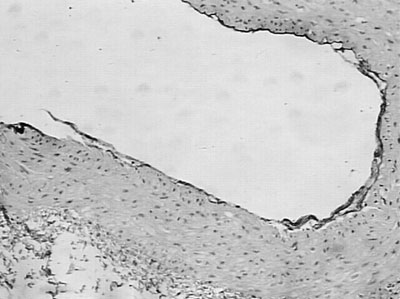
Article receive on Wednesday, March 1, 2006

CCBY All scientific articles published at bjcvs.org are licensed under a Creative Commons license

Indexes

All rights reserved 2017 / © 2024 Brazilian Society of Cardiovascular Surgery DEVELOPMENT BY