Article

lock Open Access lock Peer-Reviewed

46

Views

ORIGINAL ARTICLE

Proportion among the segments of the normal tricuspid valve annulus: parameter for valve annuloplasty

Fernando ANTONIALII; Domingo M BraileII; Glória Maria Braga POTÉRIOIII; Cledicyon Eloy da COSTAIV; Maurício Marson LopesV; Gustavo Calado de Aguiar RibeiroVI; Luciano dos Santos TARELHOVII

DOI: 10.1590/S0102-76382006000300005

INTRODUCTION

"Modern heart surgeons should pay more attention to this valve disease which is frequently ignored but kills many of our patients".

With these words, Revuelta ends his work referring to functional insufficiency of the tricuspid value, stressing the necessity of surgical repair for this valve disease [1].

Indeed, few authors have defended repair for tricuspid insufficiency associated with the surgical treatment of the mitral or aortic valves and only recently this concept has been more extensively publicized [2]. Even with the repair for valve diseases on the left, tricuspid insufficiency can persist or recur, with the necessity of further operations with high morbidity and mortality rates [3].

Tricuspid insufficiency can be caused by organic alterations of the valve or by dilatation of the ring and loss of coaptation of the cuspids. Functional tricuspid insufficiency is characterized by dilatation of the valvar annulus due to pulmonary hypertension caused by valve diseases on the left [4]. Among patients with severe stenosis or mitral valve insufficiency, from 10% to 50% have tricuspid insufficiency [5].

The treatment of functional insufficiency of the tricuspid valve by valvuloplasty is currently the most accepted technique. Thus, knowledge of the normal anatomy of the tricuspid valve and of anatomopathological changes in functional insufficiency is necessary.

It is known that dilatation of the anterior and posterior segments correspond to 5/6 of the total dilatation of the tricuspid annulus [6]. Thus, treatment of the dilatation of these segments by annuloplasty restores most of the normal anatomy of the tricuspid valve ring because the septal segment is affected very little.

Nevertheless, to correctly perform tricuspid annuloplasty an estimation of the suitable size of the valvar annulus to be used in the procedure is necessary. And so, knowledge of the proportions of the different segments of the normal tricuspid annulus is of great importance.

Although there are studies describing the anatomy of the tricuspid valve, the methodologies utilized when measuring it vary and there is no consensus in relation to the measurements taken and to the ratios among them [6-9]. Hence, the purpose of this work is to determine the ratios among the different segments that compose the normal tricuspid annulus.

METHOD

This is a descriptive study of 30 hearts from adult cadavers without fixation and that complied with the following inclusion criteria:

 The study was performed a maximum of 6 hours after death;

 Absence of congenital or acquired lesions;

 The tricuspid valves had no deformities of cuspids, chordae and papillary muscles;

 The tricuspid valves were continent.

The work was developed in the Coroner's Office in Campinas, Brazil with the approval of the local Ethics Committee.

The hearts were removed from the cadavers by technicians of the Coroner's Office by sectioning the vena cavae, pulmonary veins, aorta and pulmonary artery. After cleaning of chambers, the empty hearts were weighed. The walls of the right atrium and interatrial septum were excised between 0.5 cm and 1 cm from the atrioventricular groove and the pulmonary arteries were ligated.

Evaluation Method of the functional state of the tricuspid valves

Equipment prepared for the research consisting of three parts: a support for the heart, a perfusor and an irrigation system.

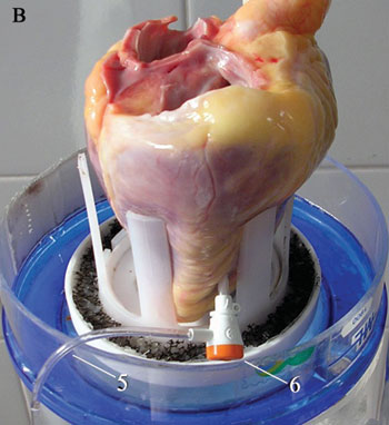
Each heart was placed on a support of parallel struts with the apex turned upside-down and the tricuspid valve easy to see. Subsequently, the apex was punctured at the right ventricle (RV) without harming the structures of subvalvar system and the perfusor was introduced into this cavity (Figure 1). A 7F cannula was utilized as the perfusor (similar to with vascular catheterism), sectioned at 3 cm of length and connected to the tubes of the irrigation system. This system comprised a water reservoir fixed to a metal support placed 50 cm above the heart. Water was infused into the RV using this system.


Fig. 1 - A) Heart in position to be submitted to evaluation. B) Side view of heart positioned in the support and with perfusor introduced into right ventricle.


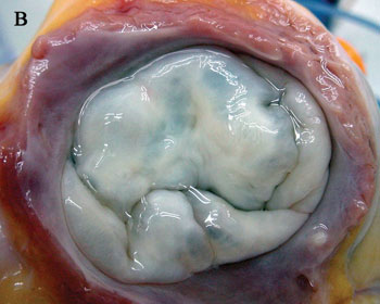
The tricuspid valves were considered continent if the RV filled with the tricuspid valve completely closed and there was no reflux of fluid and so the heart was included in the study (Figure 2). The other hearts were considered inadequate.


Fig. 2 - A) Start of the closing of tricuspid valve with infusion of water inside of right ventricle. B) Complete closure and continence of tricuspid valve.


Method to measure the valvar annulus

Measurements were obtained using a computer program and digital photographs. Photographs were taken of the tricuspid valve immediately after removing the cadaver heart. A computer analysis of the photographs was achieved utilizing the UTHSCSA program - Image Tool for Windows, version 1.28.

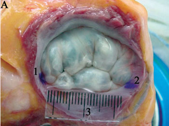
Measurements were attained at two stages: 1) with photographs of the tricuspid annulus in the anatomic position (complete valvar annulus). The anteroseptal and posteroseptal commissures were marked with water-resistant ink, according to the method described by Silver et al. [7]. Photographs were made with the RV full, coaptation of cuspids of the tricuspid valve with a millimeter ruler close to the valve (Figure 3A). The computer program was calibrated using this ruler and the following measurements were taken: perimeter of the valvar annulus, septal segment, anteroposterior segment and linear distance between the anteroseptal and posteroseptal commissures.


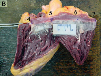
Fig. 3 - Continent tricuspid valve and with complete ring (1-Marking of the anteroseptal commissure; 2 - Marking of the posteroseptal commissure; 3 - Milimeter ruler of the same plane of the valve . B) Tricuspid valve with flattened ring (4 and 7 - Marking of the posteroseptal commissure, divided by the incision that corrects the valvar annulus; 5 - Marking of the anteroseptal commissure; 6 - Marking of the anteroposterior commissure). Presence of three millimeter rulers,from left to right, to the septal, anterior and posterior segments of the tricuspid annulus


In the second stage, measurements were made with photographs attained after the sectioning and correction of the valvar annulus (corrected valvar annulus). The sectioning was performed with an incision from the posteroseptal commissure to the apex of the RV extending towards the pulmonary valve bordering the interventricular septum. Marking of the anteroposterior commissure was achieved following the aforementioned method [7] and three millimeter rulers were used, one corresponding to each segment of the tricuspid annulus (Figure 3B). With these photographs measurements of the septal, anterior and posterior segments were made, using the computer program calibrated using the respective rules. The valvar perimeter was obtained by summing the segments.

Data in respect to age, gender, race and body surface (BS) of the cadavers were registered, as well as the weight of the heart and the cause of death.

Each computed measurement was made five times and after excluding the highest and the lowest values, the mean sizes of each segment and of the perimeter of tricuspid annulus were calculated.

From these mean values the following ratios were calculated:

 Perimeter of annulus-HS;

 Perimeter of annulus-weight of heart;

 Each segment-perimeter of annulus;

 Sum of anterior and posterior segments-perimeter of annulus;

 Sum of anterior and posterior segments-septal segment;

 Linear distance between anteroseptal and posteroseptal commissures-septal segment (ratio calculated only with the complete rings).

Comparisons between the measurements of the complete valvar ring and the flattened valvar ring were made.

Statistical analysis

The measurements are expressed as means, standard deviations, medians and 95% confidence intervals. The spearman correlation was calculated to evaluate the relationships between the measurements performed under similar conditions. Linear regression was used to create conversion formulas. Comparisons among measurements made under different conditions were achieved using the Student t-test. An ?-error of 5% was considered acceptable.

RESULTS

Characterization of the sample

Sixty-seven hearts were analysed to obtain 30 organs that complied with the inclusion criteria. Seven hearts were excluded due to anatomic alterations and 30 that demonstrated insufficiency of the tricuspid valve during the evaluation.

The mean weight of the 30 hearts was 355.5 ± 65.3 g. A total of 76.7% (23) were from men and 83.3% (25) from Caucasians. The mean age of cadavers was 43.5 ± 20.8 years and the mean body surface area was 1.83 ± 0.18 m2.

Measurements of the complete valvar ring

The mean values of the perimeter, septal and anteroposterior segments of the tricuspid annulus and the mean linear distance between the anteroseptal and posteroseptal commissures together with the results of descriptive analysis are illustrated in Table 1.


The mean values of the calculated ratios using these measurements are also shown in Table 1. There was a significant correlation between the ring perimeter and the weight of the heart (Spearman coefficient=0.4134). The ratios between different segments and between the segments with the perimeter also demonstrated significant differences giving a Spearman coefficient >0.7.

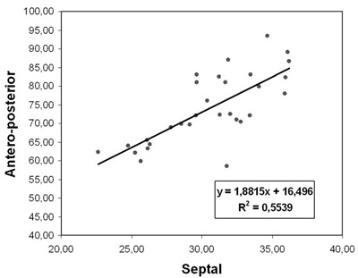
The ratio between the sizes of the septal and anteroposterior segments was 1:2.43. The conversion formula, calculated from the correlation between these measurements and by linear regression was: y = l.8815 x + 16.496 with R2 = 0.5539 (where y is the size of the anteroposterior segment and x is the size of the septal segment - Figure 4).


Fig. 4 - Sizes of anteroposterior segment and septal segment in complete tricuspid ring (in millimeters)


Measurements of the flattened valvar ring

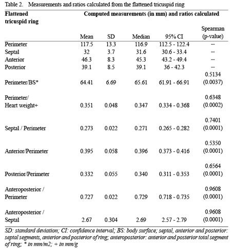
The mean sizes of the perimeter and the septal, anterior and posterior segments of the tricuspid ring together with the results of descriptive analysis are shown in Table 2.


The mean values of the ratios calculated using these measurements are also shown in Table 2. There was a significant correlation with a Spearman coefficient > 0.5 for all parameters.

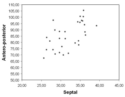
The proportions between the sizes of the septal, anterior and posterior segments of the flattened valvar ring were 1:1.45:1.22; equivalent to a ratio of 1:2.67 between septal and anteroposterior segments. The sequence of statistical tests employed showed that the relationship between measurements of the septal and anteroposterior segments is not linear (Figure 5).


Fig. 5- Sizes of anteroposterior segment and septal segment in flattened tricuspid ring (in millimeters)


Comparison between measurements obtained from the complete and flattened valvar ring.

There was a statistical difference between the sizes of segments and perimeter of the complete tricuspid valvar ring when compared to the flattened valvar ring using the Student t-test. The ratio between the anteroposterior and septal segments of the complete valvar ring (2.43) presented a statistical difference (p=0.0005) when compared to the flattened valvar ring (2.67) - Table 3.


DISCUSSION

Distortion that occurs in morphological studies with cadaver organs is related to rigor mortis. In experiments on hearts, ischemic muscle contracture can change the size and format of this organ [10,11]. In this study, in order to attenuate the effects of rigor mortis, evaluation was started immediately after removal of the organ. As the degree this influences the results is dependent of the time interval after death, only cadaver hearts in which death occurred within six hours prior to evaluation were included in the study.

Alterations in size and format of the heart also occur due to the process of fixation. Eckner et al. [11], as well as studying rigor mortis, analysed the effects of fixation using formaldehyde on the heart's dimensions. They observed that there were reductions in the dimensions independently of the method adopted when compared to hearts without fixation. However, in relation to the valvar rings, the smallest reduction, of approximately 5% to 9%, occurs in the group fixed using formaldehyde under pressure, followed by the use of formaldehyde with the ring flattened (6-20%) and, finally, the use of formaldehyde in hearts with complete rings (12%-25%). Differing percentages of reduction of valve sizes have been attributed to the use of formaldehyde by other authors under different experimental conditions [12,13]. In an attempt to find a state that was as close as possible to the physiological condition, the measurements in this study were performed on hearts without fixation and with the RV distended.

Distension of the RV using an injection of water under pressure had two goals: to determine the functional state of the tricuspid valve and to reduce the effect of rigor mortis, allowing the cuspids to take their natural position of coaptation in order to take photographs. Placing the water reservoir 50 cm above the heart was to produced a condition of hypertension (38 mmHg) in the heart chamber and to cause the valve to close.

A fact that deserves considering is related to the distribution of valves considered normal because, among the 67 hearts analyzed, only 30 presented with continent tricuspid valves. Of the 37 hearts excluded, anatomical changes were identified in cuspids and/or subvalvar system in only seven. The other hearts, although the valves were without evident organic changes, did not present with continence of the tricuspid valve. Thus, absence of any prior history of heart disease including death related to heart disease can not be considered the only indicator of normal hearts. These findings suggest a necessity of greater precautions when performing anatomical studies to describe patterns of normality.

With the methodology adopted in this study, the mean size of tricuspid valvar perimeter, calculated in valves with the complete ring, was 105 ± 12.7 mm which is a similar result to other publications [6-9,14]. For the measurements made on the complete ring, the combination of two cuspids, anterior and posterior, was considered due to difficulty to precisely identify the anteroposterior commissure. The mean value of the anteroposterior segment was 74 ± 9.4 mm and of the septal segment it was 30.6 ± 3.7 mm. With these values it is possible to calculate a ratio of 1:2.43 ± 0.212, that is, the anteroposterior segment is approximately 2.4 times larger than the septal segment.

These data differ from some published reports that show smaller ratios [6,8,9,14]. However, the majority of these works were performed with hearts fixed using formaldehyde [6,8,14] and a possible explanation for this difference would be that the greatest effect of fixing is related to the sizes of the anterior and posterior segments, as these have less collagen than the septal segment. Working with unfixed hearts, Andrade et al. [9], reported mean values that gave a ratio of 1:2.23 between the septal and anteroposterior segments, which is approximated the same as found in this study.

To better expose the tricuspid ring, its sectioning and flattening were adopted. Under these conditions, marking of the anteroposterior commissure was possible and thus, measurements of the three segments of valvar ring were obtained. With these marks, the mean values found were 32 ± 3.7 mm, 46.3 ± 8.3 mm and 39.1 ± 8.5 mm, respectively, for the septal, anterior and posterior segments of the tricuspid ring.

Most published reports on the anatomy of tricuspid valve were performed on flattened rings which were fixed using formaldehyde. This was the methodology adopted by Silver et al. [7] in a study mentioned in many other publications. The method described by these researchers was adopted in this study to mark the anteroseptal, posteroseptal and anteroposterior commissures.

The comparison between the measurements obtained in photographs of flattened valvar rings and of the complete ring gave a statistical difference in this study. This difference was verified both in the comparison of septal segments (p=0.003) and in the comparison of anteroposterior segments (p=0.0001). For the statistical study, the mean values of anterior and posterior segments, obtained with the flattened ring, were summed. Also there was a significant difference (p=0.0005) comparing the ratios between the anteroposterior and septal segments, obtained from the complete valvar ring (2.43 ± 0.212) and flattened valvar ring (2.67 ± 0.304). These data suggest that the sectioning and flattening of the tricuspid valvar ring cause changes in the sizes of the segments and consequently in the relationship that exist between them.

The utilization of digital photographs and computer programs for measurements is becoming more common [9,14,15]. In this study the same computer program was employed as the one used by Hueb et al. [15] who decisively questioned the "old" concept that the anterior segment of the mitral valve, as it is part of the fibrous skeleton of the heart, had a fixed size. The credibility of the data presented by these researchers allowed their statement to be accepted [16].

Although measurement by computer is well accepted and reproducible, during surgery this is performed manually. In respect to the functional insufficiency of the tricuspid valve, although some works have demonstrated that there is an increase in the septal segment when there is dilatation of the tricuspid valvar ring, it is known that this increase is much less than that found in the anteroposterior segment [6,14]. Thus, when the cause of valvar insufficiency is dilatation of the ring, measurement of septal segment can be a basis to estimate the ideal measurement for the anteroposterior segment and can make valvuloplasty possible.

Measurement of the septal segment of the tricuspid valve must be achieved using as parameters the anteroseptal and posteroseptal commissures. As, in surgery this measurement is normally made linearly, it was decided in this study, utilizing the marks present in photographs of these commissures on the complete valvar rings, to correlate the distance in a straight line between them with the size of septal segment. Thus, using linear regression it was possible to create a conversion formula (R2 = 0.9513):
Y = 0.8872 x + 1.7609, where y is the linear distance and x corresponds to the size of septal segment.

This formula will enable the development of scales for use in the intra-operative period that correctly correspond to the size of septal segment.

Concern with the practical application of these measurements and ratios between the different segments of tricuspid valvar ring was the greatest motivation of this study. It is known that tricuspid insufficiency is normally caused by dilation of valvar ring, that is, functional tricuspid insufficiency [4,12]. This valve disease is frequently related to valvar diseases involving the mitral or aortic valves. The repair of valve diseases on the left, without concomitant resolution of tricuspid insufficiency by surgery, has proven to be a conduct that causes higher long-term morbidity and mortality rates. This occurs due to the progression of right ventricular dysfunction and the high rate of reoperation [3].

In respect to the best manner to correct functional insufficiency of the tricuspid valve, there is a consensus about the superiority of valvuloplasty compared to valvar replacement [17]. However, this is not true in relation to valvuloplasty techniques. There are different opinions including some techniques that do not use orthesis [18,19] and others that utilize them as both rigid [20,21] or flexible [22,23] rings.

There is a trend in the literature of accepting the use of orthesis for the repair of dilatation of the anteroposterior segment as the technique with the best reproducibility, fewest complications and longer lasting [24]. What is indisputable is the necessity of surgical repair of this valve when it presents function alterations [25]. Frater [2] reported in an editorial of one of the most important journals on cardiovascular surgery, that surgeons who are indecisive about treating tricuspid insufficiency, must accept that a number of patients will need late surgeries, giving high morbidity and mortality rates.

Thus, the ratio of 1:2.43 between the septal and anteroposterior segments described in this study for complete valvar rings may be of great use in the surgical treatment of this heart valve, which is no less complex or significant than the others.

CONCLUSION

In conclusion, the experimental conditions adopted in this study, the sectioning and flattening of the tricuspid valvar ring, apart from altering the size of the segments, also alter the ratios that exist between them. The methodology applied using the complete valvar ring was considered the most correct and so, the sizes of the perimeter and the septal and anteroposterior segments are 105 ± 12.7 mm, 74 ± 9.4 mm and 30.6 ± 3.7 mm, respectively. This means that the septal segment occupies 29.5% of the tricuspid valvar ring, whilst the anteroposterior segment is 70.5%, giving a ratio of 1:2.4.

REFERENCES

1. Revuelta JM. The forgotten funcional tricuspid insufficiency: is valve repair necessary? In My Opnion. CTSNet 2003 Jun [acesso em 16 de junho de 2003]; Disponível em: URL: http://www.ctsnet.org/sections/innovation/valvetechnology/articles/article-9.html

2. Frater R. Tricuspid insufficiency. J Thorac Cardiovasc Surg. 2001;122(3):427-9.

3. King RM, Schaff HV, Danielson GK, Gersh BJ, Orszulak TA, Piehler JM et al. Surgery for tricuspid regurgitation late after mitral valve replacement. Circulation. 1984;70(3pt2):I193-7.

4. Waller BF, Howard J, Fess S. Pathology of tricuspid valve stenosis and pure tricuspid regurgitation - Part III. Clin Cardiol. 1995;18(4):225-30.

5. Kirklin JW, Pacífico AD. Surgery for acquired valvular heart disease. N Engl J Med. 1973;288(4):194-9.

6. Deloche A, Guerinon J, Fabiani JN, Morillo F, Caramanian M, Carpentier A et al. Étude anatomique des valvupathies rhumatismales tricuspidiennes. Application à l'étude des différentes valvuloplasties. Ann Chir Thorac Cardiovasc. 1973;12(4):343-9.

7. Silver MD, Lam JH, Ranganathan N, Wigle ED. Morphology of the human tricuspid valve. Circulation. 1971;43(3):333-48.

8. Jatene FB, Koike MK, Monteiro R, Veronezi SC, Magalhães MHG, Jatene AD. Avaliação anatômica da valva tricúspide. Rev Bras Cir Cardiovasc. 1992;7(1):22-7.

9. Andrade NMM, Tinois E, Vieira RW, Braile DM, Petrucci Jr O, Oliveira PPM et al. Coeficientes de proporcionalidade nas valvas atrioventriculares: estudo anatômico dos segmentos valvares em indivíduos normais. Braz J Cardiovasc Surg. 2005;20(3):255-60.

10. Curti HJ, Sanches PC, Carvalhal SS. Cardiac rigor mortis. Arq Bras Cardiol. 1985;45(6):439-46.

11. Eckner FAO, Brown BW, Overll E, Glagov S. Alteration of the gross dimensions of the heart and its structures by formalin fixation. A quantitative study. Virchows Archiv Abt A Pathol Anat. 1969;346:318-29.

12. Tei C, Pilgrim JP, Shah PM, Ormistom JA, Wong M. The tricuspid valve annulus: study of size and motion in normal subjects and in patients with tricuspid regurgitation. Circulation. 1982;66(3):665-71.

13. Ubago JL, Figueroa A, Ochoteco A, Colman T, Duran RM, Duran CG. Analysis of the amount of tricuspid valve anular dilatation required to produce functional tricuspid regurgitation. Am J Cardiol. 1983;52(1):155-8.

14. Hueb AC. Análise comparativa dos anéis valvares atrioventriculares esquerdo e direito na cardiomiopatia dilatada de diferentes etiologias: um estudo anatômico [Tese de Doutorado]. São Paulo;Universidade de São Paulo;2000.

15. Hueb AC, Jatene FB, Moreira LFP, Pomerantzeff PM, Kallás E, Oliveira SA. Ventricular remodeling and mitral valve modifications in dilated cardiomyopathy: New insights from anatomic study. J Thorac Cardiovasc Surg. 2002;124(6):1216-24.

16. McCarthy PM. Does the intertrigonal distance dilate? Never say never. J Thorac Cardiovasc Surg. 2002;124(6):1078-9.

17. Carpentier A, Deloche A, Hanania G, Forman J, Sellier P, Piwnica A et al. Surgical management of acquired tricuspid valve disease. J Thorac Cardiovasc Surg. 1974;67(1):53-65.

18. Kay JH, Maselli-Campagna G, Tsuji KK. Surgical treatment of tricuspid insufficiency. Ann Surg. 1965;162(1):53-8.

19. De Vega NG. La anuloplastia selectiva, regulable y permanente. Rev Esp Cardiol. 1972;25(6):555-6.

20. Carpentier A, Deloche A, Dauptain J, Soyer R, Bloundeau PH, Piwnica A et al. A new reconstructive operation for correction of mitral and tricuspid insufficiency. J Thorac Cardiovasc Surg. 1971;61(1):1-13.

21. Carvalho RG, Mulinari LA, Giublin PR, Mulasky JC, Silva Jr AZ; Lopes LR; et al. Emprego do anel de Gregori na posição invertida para o tratamento da insuficiência tricúspide: experiência inicial. Rev Bras Cir Cardiovasc. 1998;13(2):152-7.

22. Braile DM, Ardito RV, Pinto GH, Santos JLV, Zaiantchick M, Souza DRS et al. Plástica mitral. Rev Bras Cir Cardiovasc. 1990;2(2):86-98.

23. McCarthy JF, Cosgrove DM III. Tricuspid valve repair with the Cosgrove-Edwards Annuloplasty System. Ann Thorac Surg. 1997;64(1):267-8.

24. McCarthy PM, Bhudia SK, Rajeswaran J, Hoercher KJ, Lytle BW, Cosgrove DM et al. Tricuspid valve repair: durability and risk factors for failure. J Thorac Cardiovasc Surg. 2004;127(3):674-85.

25. Fukuda S, Song JM, Gillinov AM, McCarthy PM, Daimon M, Kongsaerepong V et al. Tricuspid valve tethering predicts residual tricuspid regurgitation after tricuspid annuloplasty. Circulation. 2005;111(8):975-9.

Article receive on Monday, May 1, 2006

CCBY All scientific articles published at bjcvs.org are licensed under a Creative Commons license

Indexes

All rights reserved 2017 / © 2024 Brazilian Society of Cardiovascular Surgery DEVELOPMENT BY