CLINICAL DATA
A female Caucasian infant of one month and seventeen days old living in São João da Boa Vista, São Paulo had been born prematurely after a normal delivery. Amniotic membrane rupture had occurred more than 24 hours previously, the meconium was thick at birth and the infant presented with whimpering and cyanosis followed by a convulsive crisis and hypoglycemia. The infant evolved with changes in the chest radiograph and was treated with crystalline penicillin and amicacin for pulmonary infection, which became systemic. Thus, prolonged broad spectrum antibiotic therapy was necessary. During breastfeeding dyspnea was noticed. The child was evaluated by a cardiologist who requested an echocardiogram that demonstrated a large mass in the right ventricle outflow tract (RVOT) as far as the pulmonary trunk (PT) with fungal characteristics. Treatment with amphotericin was initiated and there was an improvement in the clinical condition, but no changes in the echocardiographic image.
During the physical evaluation after arriving in our service, the infant was in a regular general state, dyspneic, ruddy, hydrated and afebrile. The heartbeat was regular with two clicks and normal sounds without murmurs. Auscultation of the lungs was symmetrical without adventitious noises. The abdomen was swollen with the liver 4 cm from the right costal border. The pulses were symmetrical and the peripheral oxygen saturation was 96%.
ELECTROCARDIOGRAM
The electrocardiogram evidenced sinusal rhythm with a frequency of 125 bpm. The QRS angle was + 60º and the PR interval was 0.16 s. The QRS was 0.08s without direct signs of right ventricular overload.
RADIOGRAM
The radiogram demonstrated visceral situs solitus. The heart area and lungs were normal.
ECHOCARDIOGRAPH
The echocardiograph showed situs solitus and levocardia with normal venoatrial, atrioventricular and ventriculo-arterial connections. The longilinea image was irregular projecting from the RVOT through the pulmonary valve to the pulmonary trunk with an irregular aspect suggestive of fungal vegetation. The infant had a good ventricular function without pericardial effusions (Figure 1).
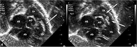
Fig. 1 - Coronal subcostal echocardiographic slices demonstrating: A - In diastole, a large vegetation in the right ventricle outflow tract (arrow). B - In systole, vegetation through the pulmonary valve extending as far as the bifurcation of the pulmonary arteries (arrows).
AE: Left atrium; AD: right atrium; VD: right ventricle; AO: aorta; TP: pulmonary trunk
The clinical history and the long period using a central nervous catheter to administer endovenous antibiotics due to neonatal sepsis, the echocardiographic and the clinical improvement with antifungal agents made the diagnosis of fungal endocarditis caused by vegetation obvious; there was a high possibility of thrombogenic events.
OPERATION
A median transsternal thoracotomy was performed and a cardiopulmonary bypass was established with hypothermia at 34 ºC. Intermittent anterograde sanguineous cardioplegia at 4 ºC was also necessary. After opening the pulmonary trunk, a large vegetation was observed originating in the RVOT. There were no adherences in the pulmonary trunk or on the pulmonary valve; the vegetation was only connected inside the RVOT which facilitated resection without damaging the pulmonary valve (Figure 2). The perfusion and myocardial ischemia times were 38 and 28 minutes, respectively. The collected material proved to be contaminated with gram positive cocos, gram negative cocos as well as numerous hyaline hyphas. In the postoperative period the patient continued to receive specific antibiotic and antifungal therapy and was released in excellent clinical conditions after 28 days. A late echocardiogram showed that the right ventricle was clear of vegetation and the heart structures were normal.

Fig. 2 - Pulmonary trunk opened lengthwise showing the large vegetation fixed in the right ventricle outflow tract extending through the pulmonary valve as far as the bifurcation of the pulmonary arteries
 All scientific articles published at bjcvs.org are licensed under a Creative Commons license


 Read in Portuguese
 Portuguese PDF
 Print
Print
 Send this article by email
 How to cite this article
 Submit a comment
 Mendeley
 Pocket