CLINICAL DATA
A six-month old female infant suffered with effort on breast feeding since three months old. She was in a regular general state, discolored +4, acyanotic, with slight tachydyspnea. Auscultation was rhythmic with a third sound, systolic murmur of +++/6 focused at the mitral valve transmitted to the axilla. The lungs presented with stertor crackles at the bases. The liver was 4 cm from the right costal border. The arterial pressure was 90/50 mmHg and the peripheral pulses of all four limbs were palpable.
ELECTROCARDIOGRAM
Sinusal rhythm was evidenced with 150 beats per minute. QRS complex axis was 0º. Q wave was deep at D1 and AVL indicating an electrically inactive area high on the lateral wall. The ST segment was high at V1, V2 and V3 suggesting tendency of injury in the anteroseptal wall and inversion of the T wave at D1, AVL, V5 and V6 demonstrated subendocardial ischemia.
RADIOGRAM
The cardiothoracic index was 0.69 with a generalized increase in the heart area. Pulmonary parenchyma with bilateral congestion was seen compatible with venocapillary hypertension.
ECHOCARDIOGRAM
The echocardiogram evidenced situs solitus at levocardia, with concordant venoatrial, atrioventricular and ventriculoarterial connections. Doppler echocardiography evidenced a significant dilation of the left ventricle with a diastolic diameter of 44 mm, significant diffuse hyperkinesias and a shortening fraction of 11.4%. Significant mitral insufficiency was also seen. The right coronary artery was dilated and anomalous origin of the left coronary artery from the pulmonary branch with retrograde blood flow.
DIAGNOSIS
A coronary cineangiography confirmed the echocardiographic findings, defining an anomalous origin of the left coronary ostium deriving from the pulmonary branch. The right coronary artery was dilated and supplied the complete left coronary system by a retrograde flow.
OPERATION
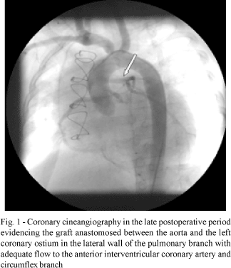
Median transsternal thoracotomy was performed and cardiopulmonary bypass was established. The correction was performed under hypothermia of 18 ºC using intermittent anterograde sanguineous cardioplegia at 4 ºC. The left coronary ostium originated in the lateral wall of the pulmonary branch. The left subclavian artery was resected as a free graft. The pulmonary branch was transversally opened and anastomosis of the left subclavian artery was performed between the aorta and the left coronary ostium similar to the technique described by Takeuchi [1]. With the aim of avoiding pulmonary supravalvar obstruction, the transversal incision of the pulmonary branch was enlarged with a bovine pericardial patch. The times of perfusion and myocardial ischemia were 213 and 140 minutes respectively. The patient evolved with significant ventricular dysfunction and low outflow syndrome in the immediate postoperative period, requiring 12 days in the intensive care unit with intravenous inotropic agents. She was released from hospital after 32 days taking acetylsalicylic acid, digital and diuretics. In the fourth postoperative month presented with class IV heart insufficiency (NYHA), with a suspicion of graft occlusion. A coronary cineangiography was performed which evidenced a normal graft. An echocardiography demonstrated a reduction in the diameters of the left ventricle and improvement of the contractibility.
BIBLIOGRAPHIC REFERENCE
1. Takeuchi S, Imamura H, Katsumoto J, Hayashi I, Katohgi T, Yozu R et al. New surgical method for repair of anomalous left coronary artery from the pulmonary artery. J Thorac Cardiovasc Surg 1979;78:7-11.

 All scientific articles published at bjcvs.org are licensed under a Creative Commons license